Standing Calf Raise Foot Positioning in Rehabilitation
May 06, 2022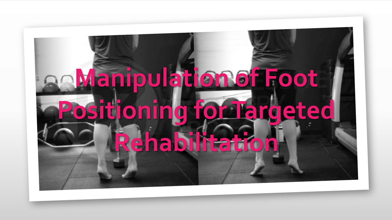
Manipulation of Foot Positioning for Targeted Rehabilitation
When it comes to rehabilitation, we know that WHAT you are doing with your exercise prescription will yield specific tissue adaptations, but also HOW you perform your exercise can broaden the scope even more.
Today I will focus on the effects of foot positioning on the standing calf raise exercise.
In this instance the WHAT refers to our 'dosage'.
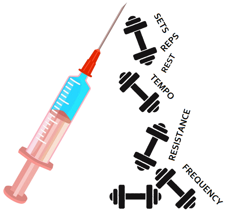
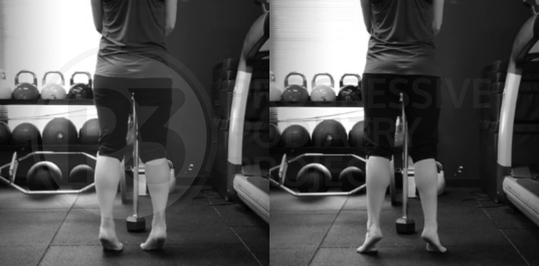
The HOW refers to our positioning, and in this instance we will explore how an abducted or adducted foot position may alter the tissue responses when performing the standing calf raise.
Given the function of the gastroc-soleus complex, not to mention its involvement in a large array of lower extremity musculoskeletal pathologies (either in the cause or effect role - or both!) it's no wonder the calf raise exercise presents such a large clinical utility for practitioners in a rehabilitation & performance setting.
When we have an understanding of the ways in which we can manipulate dosage variables for the calf raise, this exercise can give you even more bang for your buck!
Effects of Foot Positioning
A number of studies have investigated various metrics and subsequent adaptations from performing the calf raise exercise in neutral, abducted and adducted positions.
Typically the variations are performed with the feet roughly 30 deg ABducted or 30 deg ADducted. However, when it comes to prescribing these variations, go with what is comfortable for the client. That is, if they can't comfortably perform the exercise 30 degrees adducted but 10-15 degrees works for them, then that's the position to go with.

EMG Activity
Akuzawa et al (2017) & Reimann et al (2011) have published findings regarding the EMG activation of lower extremity muscles in relation to foot positioning.
Standing calf raises performed using a neutral stance elicited similar levels of medial and lateral gastrocnemius EMG activation, during both the concentric and eccentric phases of the exercise.
Differences in EMG activity of a number of lower extremity muscles were found to occur throughout both the concentric & eccentric phases of the standing calf raise, with the feet in either abducted or adducted positions (table summarises the findings below).
Feet in an abducted position significantly increase medial gastrocnemius EMG activity compared to lateral gastrocnemius.
Feet in an adducted position significantly increase lateral gastrocnemius EMG activity compared to medial gastrocnemius.
Hypertrophy
Nunes et al (2020) found that foot positioning produced varying hypertrophic adaptations of the medial & lateral heads of gastrocnemious.
Whilst all foot positions resulted in increases in muscle thickness (hypertrophy), increases varied between medial & lateral heads as a result of foot adduction or abduction.

As there are a limited number of studies in relation to foot positioning and the subsequent influences on adaptations, future work is needed to further test these findings.
However, as clinicians we can use this information to assist our clinical reasoning when it comes to exercise prescription and its role in achieving our therapeutic goals.
Why Would Foot Position Alter Responses?
When we take the above information and combine it with what we know regarding how tissues respond to load, it can help us understand why this may be occurring.
For instance, when it comes to triggering adaptations within muscle & tendon tissue, we know that there can be differences in the expression & responses of various proteins and enzymes between eccentric and concentric contractions.
For example: eccentric contractions result in higher rates of Type I & Type III Collagen synthesis in the gastrocnemius muscle compared to concentric contractions (Heinemeier et al, 2007)
If we couple the above with further manipulating foot position, it stands to reason that by altering foot positioning we are altering the lever arm of the calf muscle complex in relation to the ankle joint.
This will result in either ↑ or ↓ the lever arm of the medial/lateral heads of the gastrocnemius, in relation to either an abducted or adducted position of the foot.
Thereby creating an increased range of movement when moving the ankle into a more dorsiflexed position, subsequently increasing eccentric loading.
Positioning for Targeted Pathology Rehabilitation
A skill required of healthcare practitioners is being able to apply the evidence in a clinical setting, in relation to the individual who is seeking our care.
By understanding the effects the movement variations will have on loading various connective tissues, we can become more targeted in our rehabilitation programming.
This doesn't mean we should be aiming to over-complicate rehabilitation programming at all. It simply means applying sound clinical reasoning to achieve our therapeutic goals, so we can help our clients achieve their treatment goals.
A great example of the clinical utility of foot manipulation of the standing calf raise, may be in the rehabilitation of a medial gastrocnemius strain at the myotendinous junction.
We know that strain injuries most commonly occur as a result of excessive active loading, more specifically, excessive active lengthening. The majority of the time (but not always) lengthening alone is not sufficient to result in a strain injury, it almost always requires muscle unit activation.
Eccentric muscle contractions serve to decelerate the body &/or to store elastic recoil energy.
If an individual experiences a calf strain injury to the medial gastrocnemius myotendinous junction, we may wish to target rehabilitation in this area.
In the early stages of healing we may not want a greater degree of eccentric load on the area, so an adducted calf raise may serve to introduce a lower degree of eccentric load to the injury site compared to the lateral head of the gastrocnemius.
As we build capacity and positive adaptation is occurring, we may move the foot to neutral and then into abduction, where we will see greater degrees of eccentric strain loads being placed upon the medial head of the gastrocnemius.
Throughout this process we may also be altering other dosage variables to further accentuate tissue responses to achieve our therapeutic goals.
Foot Position
+
Dosage
=
Targeted Response
Q: What Other Pathologies Can Benefit From Targeted Rehabilitation?
A: Lots
The purpose of these articles is to share knowledge, but also serve to help you build you clinical and critical thinking skills.
In saying that, I won't be spoon feeding all of the answers to you today!
I'd like your input. What are some other pathologies in which you believe manipulating foot positioning (+/- prescription dosage) could be beneficial?
To help you answer this questions, here are some questions we need to ask ourselves when we are prescribing an exercise in order to achieve a desired tissue response.
- What is the therapeutic goal?
- What adaptations need to occur in order to achieve the therapeutic goal?
- What structures require adaptations in order to achieve the therapeutic goal?
- What movements will allow the identified structures to be loaded in a way that will result in the desired adaptations?
- When the foot is in an ABducted position - what tissues/structures may have ↑ loads?
- When the foot is in an ADducted position - what tissues/structures may have ↑ loads?
Loads in this instance don't have to be purely eccentric/concentric, not do they have to be specific to loading muscle tissue, think of tensile & compressive loads to other connective tissues as well (there are a multitude of structures that surround the ankle joint).
Leave your answers below!
Are you a health practitioner who would like to improve your exercise prescription outcomes?
The Movement Prescription Blueprint is designed to help you do just that!
The MPB will help guide you through the exercise prescription process, allowing you to explore the 3 KEY elements of exercise prescription with your client, to help set you (and your clients) up for treatment success!
To download my FREE resource, The Movement Prescription Blueprint click HERE

References:
Akuzawa, H., Imai, A., Iizuka, S., Matsunaga, N., & Kaneoka, K. (2017). The influence of foot position on lower leg muscle activity during a heel raise exercise measured with fine-wire and surface EMG. Physical Therapy In Sport, 28, 23-28. doi: 10.1016/j.ptsp.2017.08.077
Heinemeier, K., Olesen, J., Haddad, F., Langberg, H., Kjaer, M., Baldwin, K., & Schjerling, P. (2007). Expression of collagen and related growth factors in rat tendon and skeletal muscle in response to specific contraction types. The Journal Of Physiology, 582(3), 1303-1316. doi: 10.1113/jphysiol.2007.127639
Nunes, J., Costa, B., Kassiano, W., Kunevaliki, G., Castro-e-Souza, P., & Rodacki, A. et al. (2020). Different Foot Positioning During Calf Training to Induce Portion-Specific Gastrocnemius Muscle Hypertrophy. Journal Of Strength And Conditioning Research, 34(8), 2347-2351. doi: 10.1519/jsc.0000000000003674
Riemann BL, Limbaugh GK, Eitner JD, LeFavi RG. Medial and lateral gastrocnemius activation differences during heel-raise exercise with three different foot positions. J Strength Cond Res. 2011 Mar;25(3):634-9. doi: 10.1519/JSC.0b013e3181cc22b8. PMID: 20581696.
Join the Research Round-Up
Free monthly newsletter, delivering the latest research straight to your inbox
Research Round-Up delivered at the end of each month