
The Supination Resistance Test - What, Why & How...
Feb 20, 2024
Recently I shared some insights on the clinical utility of the Foot Posture Index (FPI) - find the podcast episode here.
Within this episode I discussed how I find the FPI and Supination Resitance Test useful when it comes to making footwear recommendations, as well as identifying some appropriate orthoses prescription variables to include in a script.
Today, I want to dive a little deeper into the Supination Resistance Test.
WHAT it is, WHY we would potentially use it, and HOW to perform the test.
What is the Supination Resistance Test?
The Supination Resistance Test (SRT) is a clinical (and now research) tool developed by Dr Kevin Kirby, that is used to assess the amount of external force required to initiate supination of the foot.
It provides clinicians with a degree of clinical utility relating to the prescription of foot orthoses, especially when it comes to understanding what script variables we may need to manipulate to alter mechanical loading of the lower extremity.
Essentially, the supination resistance test helps determine the force, often expressed in Newtons of force (N), needed to supinate the foot.
"A greater supination resistance may indicate greater loads on structures responsible for generating internal supination moments across the subtalar joint during static and dynamic tasks."
Greater supination resistance may be an expected finding in medial foot and ankle musculoskeletal disorders, such as plantar fasciopathy (PF) and posterior tibial tendon dysfunction (PTTD).
Reduced supination resistance may be present in lateral ankle disorders, such as chronic ankle instability (CAI)."
Moisan et al (2023)
There are two common methods used to test supination resistance.
1) The Manual Supination Resistance Test
2) The validated Keystone device
Both body weight and subtalar joint axis are related to supination resistance (6).
Given how both the manual and device tests are performed, it's entirely reasonable that body weight plays into the equation.
In regards to the subtalar joint axis, it was reported that the greater the perpendicular distance from the fifth metatarsal head to the subtalar joint axis resulted in a higher degree of supination resistance (6).

My best attempt at creating image to demonstrate STJ axis & 5th metatarsal head position relative to medially deviated STJa (left), "normal" STJa (middle), laterally deviated STJa (right)
Performing the Manual Supination Resistance Test
(Griffiths & McEwan, 2012)
Instructions
Instruct person to stand barefoot in their relaxed calcaneal stance position (RCSP) and adopt a natural angle and base of gait.
Places the tips of your index and middle fingers directly plantar to the medial aspect of the navicular and then pull upward, in a vertical plane and parallel to the tibia.
Clinician rates how the foot responds to the force and subjectively rates the resistance to supination on a scale from 0 to 5.
0 = very low resistance to supination → 5 = very high resistance to supination.
 Manual Supination Resistance Test (Image © T.Reeve)
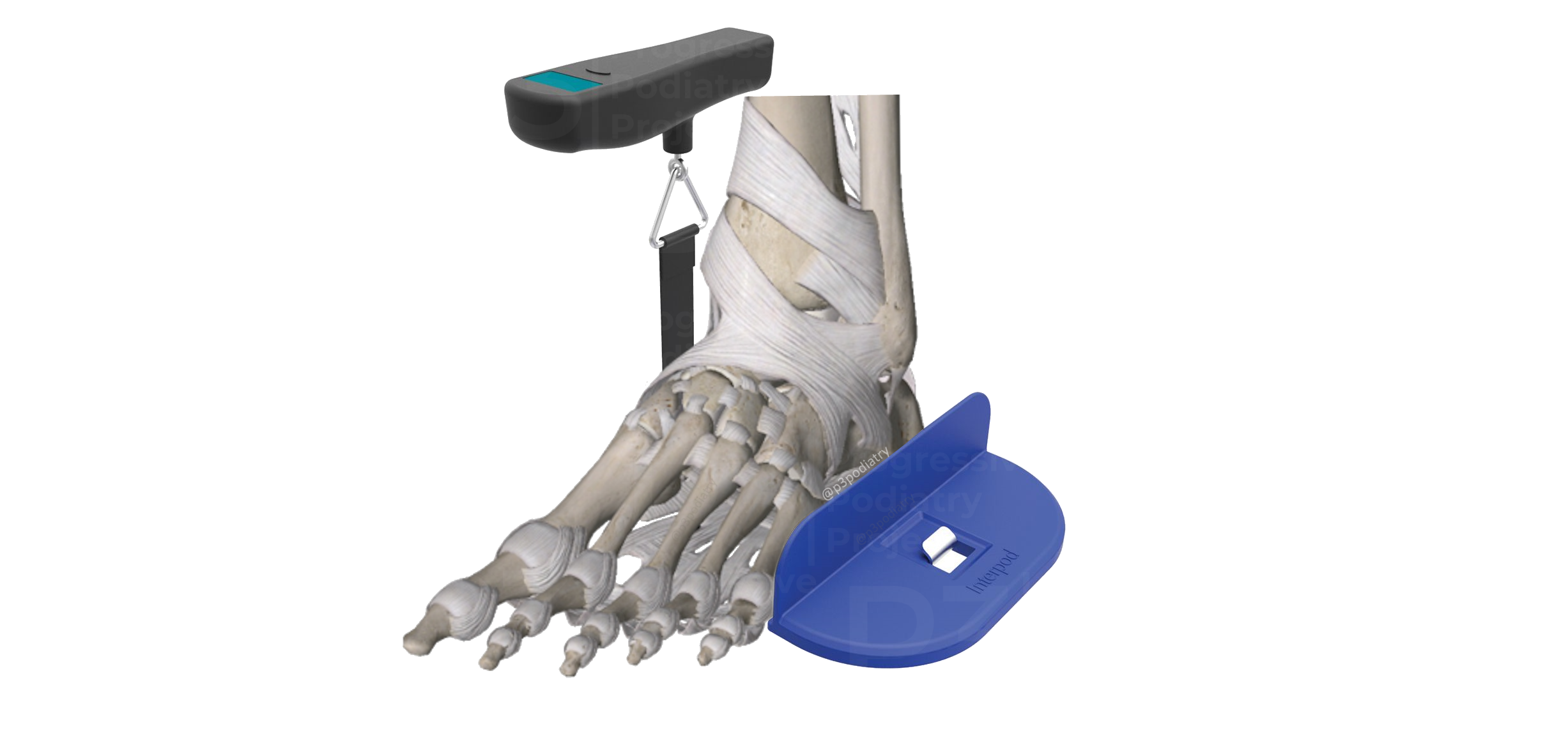
Performing the SRT Using the Keystone Device
(Moisan et al, 2023 & Moisan et al, 2021)
Prior to Performing Test
The individual's height, mass, age, and Foot Posture Index (FPI) score are recorded before the test.
Individual is instructed to march in place for five steps and then come to rest in their resting calcaneal stance position (RCSP).
Clinician marks a vertical line on the posterior calcaneus to assist in visualising calcaneal inversion.
Device Setup
Attach a 25-mm-wide non-stretchable strap to an anchor on the ground, positioning it beneath the foot by gently lifting the heel of the foot to be tested.
Adjust the strap so that it passes from the calcaneocuboid region laterally to just posterior to the navicular tuberosity medially.
Testing Procedure
Instruct the participant to stand upright with weight evenly distributed between the feet and look straight ahead during testing.
Testing Execution
Apply vertical traction to the strap (pull up on the device) at a constant speed, until just enough force is applied to initiate inversion motion of the calcaneus.
Ensure the individual does not assist (pull upward) or resist (push downward) the pull of the tester during the test.
Verify there is no active muscular activity, including contraction of the anterior tibialis muscle.
 Keystone image sourced from footmedics.com.au - final image created by © T.Reeve)
Reliability & Validity
Moisan et al (2021) assessed the reliability of the Keystone device and reported that both interrater and intrarater reliability scores were good.
Relationship Between Supination Resistance, Kinematics & Kinematics
McBride et al (2019)
The SRT, when performed using the Keystone device, is reliable and related to midfoot kinetics during gait (4).
Kinetics and kinematics are two fundamental concepts in the field of biomechanics that are used to study the motion of objects, including the human body.
Kinematics
Kinematics is the branch of mechanics that deals with the motion of objects without considering the forces causing the motion.
It focuses on describing the spatial and temporal aspects of motion, such as position, velocity, acceleration, and the trajectory of movement.
In human movement analysis, kinematics describes how body segments move relative to each other without considering the forces that produce the motion.
Examples of kinematic measurements include joint angles, segmental positions, and movement patterns.
Example
Analysing the hip, knee, and ankle joint angles during the gait cycle.
Kinetics
Kinetics is the branch of mechanics that deals with the forces acting on objects that cause motion.
It involves the study of the forces, torques, and moments that produce or change the motion of an object.
In human movement analysis, kinetics focuses on understanding the forces generated by muscles, tendons, ligaments, and external factors that influence movement.
Examples of kinetic measurements include joint moments, ground reaction forces, muscle forces, and energy transfer during movement.
Example
The measurement of ground reaction forces (GRF) or power generation of a muscle during activities like running or jumping.
"In this study, the results of the supination resistance test were associated with peak midfoot pronation moments, but not peak ankle inversion moments.
Furthermore, moderate to good significant relationships were found with peak midfoot plantarflexion moments and midfoot power generation."
Midfoot Pronation Moments
The study identified an inverse relationship between supination resistance measured in the Relaxed Calcaneal Stance Position (RCSP) and midfoot pronation moments during gait.
This suggests that as supination resistance increases, midfoot pronation moments decrease.
"We suspect that the inverse relationship between the SRT in RCSP and midfoot pronation moments occurs because the SRT measures the ability of the forefoot to supinate on the hindfoot.
As less force is needed for forefoot supination to occur, greater control may be required from the midfoot pronators to coordinate the amount of supination that takes place in late stance."
Peak Midfoot Plantarflexion Moments
The SRT demonstrated significant relationships with peak midfoot plantarflexion moments during gait.
This indicates that supination resistance may influence the generation of plantarflexion moments in the midfoot region.
Peak Midfoot Power Generation
Relationships were observed between the SRT and peak midfoot power generation during gait.
This suggests that supination resistance may be linked to the generation of power in the midfoot area.
Midfoot power generation relates to the production of mechanical energy by the midfoot joints and structures (muscles, tendons etc) as part of the overall propulsion and support mechanism of the foot during walking or running.
Q: Why don't we just rely on the FPI-6 (& other similar measures)?
A: Static measures often don't correlate with dynamic function.
A pronated foot (per FPI-6) may have low supination resistance.
A "Neutral" foot (per FPI-6) may have high supination resistance.
Supination Resistance in Lower Extremity Musculoskeletal Pathologies
A number of years ago I when I started using the Keystone device, I came across a number of articles by Craig Payne over at http://www.runresearchjunkie.com/
Some of these I'm not certain on if they ever made it to research publication or not (as the primary data, not just secondary).
However, within his posts Craig discusses a number of pathologies in which he has measured the supination resistance for.
Keep in mind that we still do not have normative values for the SRT and Craig mentions within these posts that the information/numbers provided are secondary data from another project - so he did not recommended to relying on this too heavily.
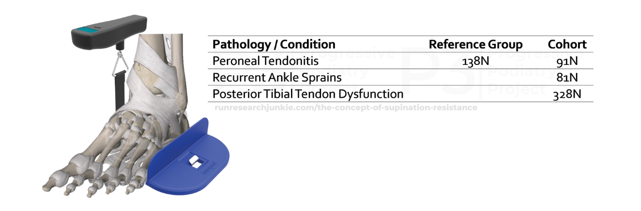
Summary of findings from runresearchjunkie.com/the-concept-of-supination-resistance
Fast forward to today, where we saw a paper published quite recently that despite having different data representation (N vs %BW), we now have two papers demonstrating some relative consistency.
Supination Resistance Variations in Foot and Ankle Musculoskeletal Disorders: Implications for Diagnosis and Customised Interventions with Wedged Insoles.
Moisan et al (2023)
The objectives of this study were to;
-
Determine if supination resistance differs across musculoskeletal disorders (CAI, PF, PTTD) compared to controls.
-
Identify the differences in supination resistance between injured and healthy feet.
-
Investigate the changes in supination resistance observed in individuals with musculoskeletal disorders after simulating full-length varus and valgus wedging of the foot.
56 participants were assessed (n = 14 for each - CAI, PF, PTTD & healthy controls)

Results table from Moisan et al (2023) - open access
Results
Individuals with chronic ankle instability (CAI) exhibited lower supination resistance in the injured foot compared to the healthy foot.
Those with posterior tibial tendon dysfunction (PTTD) showed greater supination resistance in the injured foot.
Plantar fasciopathy (PF) did not show significant differences in supination resistance between injured and healthy feet.
Varus and valgus inclinations to the surface were effective in modifying supination resistance in PTTD and CAI, respectively.
The use of foot orthoses or insoles with varus or valgus posting may be beneficial for patients with PTTD and CAI, respectively.
In all studies that I have found and included relating to supination resistance, the authors consistently state that more trials aiming to investigate the relationship between specific lower extremity musculoskeletal pathologies & supination resistance are required.
Clinical Utility
Based on the findings of these papers, plus a pinch of clinical experience, when it comes to assessing and managing a number of lower extremity pathologies (CAI & PTTD especially), subtalar joint axis and supination resistance are two measures we should consider including in our biomechanical assessments for lower extremity musculoskeletal pathologies, as we can gather useful information that may influence;
-
Our footwear recommendations (ie: suitable characteristics).
There are a number of footwear construction elements that influence the fit, feel & function of a shoe.
Free downloadable footwear prescription resource → The Footwear Prescription Blueprint (get it here) -
Our orthomechanical prescriptions (ie: orthoses script).
Especially our hindfoot prescription variables. -
Suitability of exercise therapies (will explain another time).
I hope this article has shed some light on the Supination Resistance Test for you and provided some clarity on when we may utilise it.
As always, I'd love to hear your thoughts.
Do you assess subtalar joint axis &/or supination resistance regularly in your biomechanical assessments?
If you do, how does it inform your practice?
If you don't, is there a reason for not conducting it?
Keep an eye out on this blog, as I am in the process of putting together another article to expand on some orthoses script components that I may consider for a low/high supination resistance & medially/laterally deviated subtalar joint axis (this will be a few weeks off as I am currently drowning in a workload of my own creation).
References
-
Bennett, P. J., Lentakis, E., & Cuesta‐Vargas, A. (2015). Limitations of the manual supination Resistance Test. Journal of Foot and Ankle Research, 8(S2). https://doi.org/10.1186/1757-1146-8-s2-o2
-
Griffiths, I., & McEwan, I. (2012). Reliability of a new supination resistance measurement device and validation of the manual supination Resistance Test. Journal of the American Podiatric Medical Association, 102(4), 278–289. https://doi.org/10.7547/1020278
-
McBride, S., Dixon, P., Mokha, M., & Samuel Cheng, M. (2019). The relationship between supination resistance and the kinetics and kinematics of the foot and ankle during gait. Gait & Posture, 73, 239–245. https://doi.org/10.1016/j.gaitpost.2019.07.305
-
Moisan, G., Chicoine, D., McBride, S., Farahpour, N., Isabelle, P., Dagenais, C., & Griffiths, I. (2023). Supination resistance variations in foot and ankle musculoskeletal disorders: Implications for diagnosis and customised interventions with wedged insoles. Journal of Foot and Ankle Research, 16(1). https://doi.org/10.1186/s13047-023-00681-5
-
Moisan, G., McBride, S., Isabelle, P., & Chicoine, D. (2021). The keystone device as a clinical tool for measuring the supination resistance of the foot: A reliability study. Musculoskeletal Care, 20(3), 570–576. https://doi.org/10.1002/msc.1614
-
Payne, C., Munteanu, S., & Miller, K. (2003). Position of the subtalar joint axis and resistance of the Rearfoot to supination. Journal of the American Podiatric Medical Association, 93(2), 131–135. https://doi.org/10.7547/87507315-93-2-131
-
Payne, C. (2013). The concept of ‘Supination Resistance’. Running Research Junkie. http://www.runresearchjunkie.com/the-concept-of-supination-resistance
Join the Research Round-Up
Free monthly newsletter, delivering the latest research straight to your inbox
Research Round-Up delivered at the end of each month